×
模态框(Modal)标题
在这里添加一些文本
Close
Close
Submit
Cancel
Confirm
×
模态框(Modal)标题
×
Chinese Medical E-ournals Database
Adv Search
Fig/Tab
导航切换
中华妇幼临床医学杂志(电子版)
Home
Just Accepted
Current Issue
Archive
Video
About Journal
Publication Policies
Aims & Scope
Indexed-in
Editorial Board
中文
Figure/Table detail
Liquid-Based Cervical Cytology for Cervical Cancer Screening: A Retrospective Analysis of 61610 Cases
Ai-guo MA, Hui-juan LU
Chinese Journal of Obstetrics & Gynecology and Pediatrics(Electronic Edition)
, 2012, 08(
01
): 21-27. DOI:
10.3877/cma.j.issn.1673-5250.2012.01.006
Figure 3B
Follow up histology of the patient of the left figure was invasive endocervical adenocarcinoma. (HE ×100)
Other figure/table from this article
Table 1
Primary and re-review interpretation of 61 610 cases by liquid-based cytology test [
n
(%)]
Table 2
Interpretation of cytology versus reference standard (histology diagnosis)
Table 3
Interpretation of conventional Pap smears versus reference standard (histology diagnosis)
Table 4
Detection of squamous intraepithelial lesion and squamous cell carcinoma by both cytological techniques versus reference standard (histology diagnosis)
Figure 4B
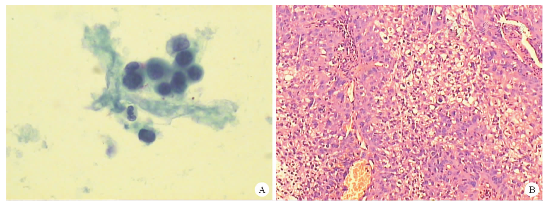
Follow up histology was invasive nonkeratinized small-sized squamous carcinoma of the cervix. (HE ×100)
Figure 5B
Follow up histology of the patient of the left figure was invasive nonkeratinized small-sized squamous carcinoma of the cervix. (HE ×100)
Figure 1B
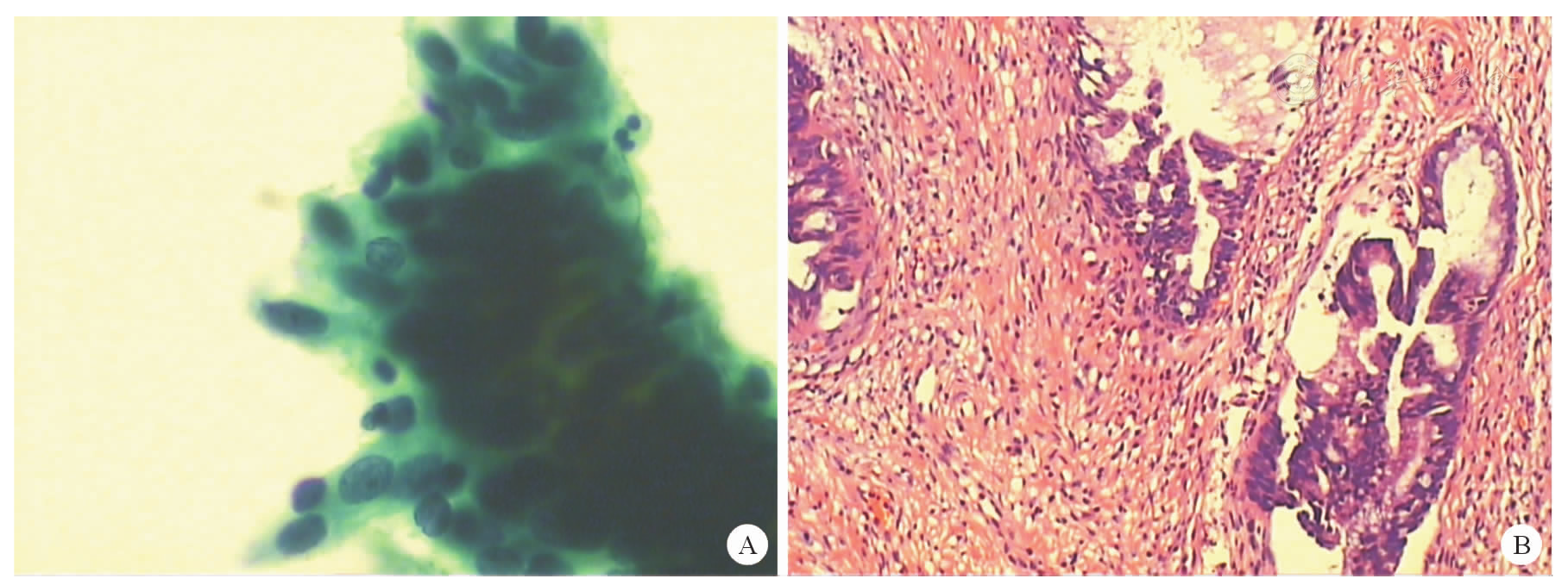
Follow up histology the patient of the left figure was cervical superficial adenocarcinoma with foci AIS. (HE ×100)
Figure 2B
Follow up histology of the patient of the left figure was AIS. (HE ×100)
Copyright © Chinese Medical Association, Chinese Medical Multimedia Press 京ICP备14006079号-1 网络出版服务许可证:(署)网出证(京)字第075号