×
模态框(Modal)标题
在这里添加一些文本
Close
Close
Submit
Cancel
Confirm
×
模态框(Modal)标题
×
Chinese Medical E-ournals Database
Adv Search
Fig/Tab
导航切换
中华妇幼临床医学杂志(电子版)
Home
Just Accepted
Current Issue
Archive
Video
About Journal
Publication Policies
Aims & Scope
Indexed-in
Editorial Board
中文
Figure/Table detail
Liquid-Based Cervical Cytology for Cervical Cancer Screening: A Retrospective Analysis of 61610 Cases
Ai-guo MA, Hui-juan LU
Chinese Journal of Obstetrics & Gynecology and Pediatrics(Electronic Edition)
, 2012, 08(
01
): 21-27. DOI:
10.3877/cma.j.issn.1673-5250.2012.01.006
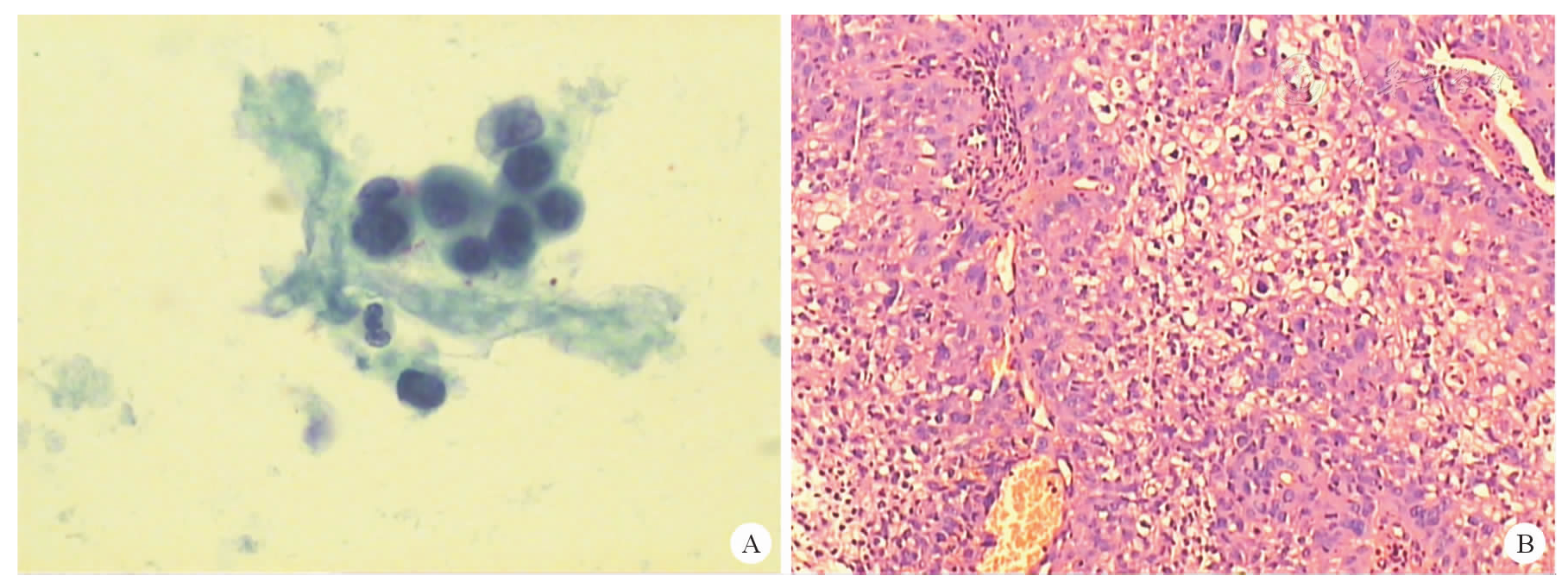
Figure 4B
Follow up histology was invasive nonkeratinized small-sized squamous carcinoma of the cervix. (HE ×100)
Other figure/table from this article
Table 1
Primary and re-review interpretation of 61 610 cases by liquid-based cytology test [
n
(%)]
Table 2
Interpretation of cytology versus reference standard (histology diagnosis)
Table 3
Interpretation of conventional Pap smears versus reference standard (histology diagnosis)
Table 4
Detection of squamous intraepithelial lesion and squamous cell carcinoma by both cytological techniques versus reference standard (histology diagnosis)
Figure 5B
Follow up histology of the patient of the left figure was invasive nonkeratinized small-sized squamous carcinoma of the cervix. (HE ×100)
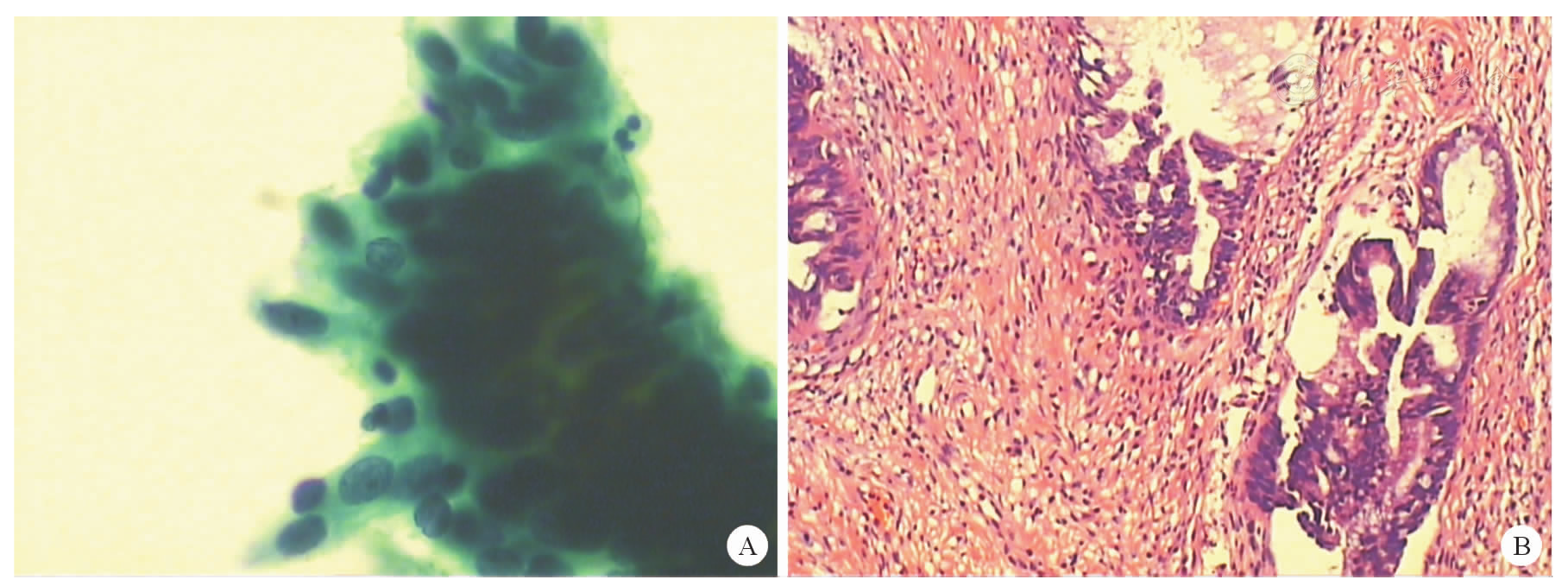
Figure 1B
Follow up histology the patient of the left figure was cervical superficial adenocarcinoma with foci AIS. (HE ×100)
Figure 2B
Follow up histology of the patient of the left figure was AIS. (HE ×100)
Figure 3B
Follow up histology of the patient of the left figure was invasive endocervical adenocarcinoma. (HE ×100)
Copyright © Chinese Medical Association, Chinese Medical Multimedia Press 京ICP备14006079号-1 网络出版服务许可证:(署)网出证(京)字第075号